


Over 20,000 spectral scans, and more than 4,500 skin lesions

DermaSensor combines advanced spectroscopy and machine learning algorithms to help clinicians evaluate skin lesions quickly, non-invasively, and objectively.
A few words from the organizations we’ve supported across strategy, operations, and digital transformation.
Trustindex verifies that the original source of the review is Google. Still in the process of getting laser tattoo removal here. I was afraid at first because I’ve never had it done but they have done such a great job. First of all, I’ve never met such kind people before. They have a great yet professional sense of humor, they always remember who I am, what I do for work, and ask about recent happenings. Their equipment always seems to be clean. These people are very knowledgeable and detail oriented. They have never messed up anything on me and they also take progress pictures so that I can see how much my tattoos have faded, which I greatly appreciate. They informed me on caring for my skin to prevent scaring and blistering from the laser. I’ve not seen any! If you are a little scared they have stress balls on hand which I have used and find very considerate and helpful :) If you need to get tattoos removed this is the place to go!Posted on GoogleTrustindex verifies that the original source of the review is Google. Showing great resultsPosted on GoogleTrustindex verifies that the original source of the review is Google. If you are looking for a physician who truly puts patients first — look no further than Dr. Rettenmaier at No Sweat. He gave me over two hours of his time, genuinely listened, and treated me with the kind of care I haven’t experienced in a long time. As someone with 33 years in critical care nursing, I recognize exceptional clinical skill when I see it. Dr. Rettenmaier is the real deal. He constantly attends conferences and stays ahead of the latest treatments so his patients have access to technology that nobody else in Indiana offers — including a machine sourced directly from South Korea that he brought here specifically for his patients. His staff is equally exceptional — talented, warm, and welcoming from the moment you walk in. You can tell a great deal about a physician by the team he builds around him, and Dr. Rettenmaier’s team reflects his same standard of excellence and genuine care for every patient. He doesn’t just treat patients. He advocates for them. If you want a provider who does what is right — not what is easy — this is your doctor.Posted on GoogleTrustindex verifies that the original source of the review is Google. No sweat made my son’s MiraDry procedure easy and comfortable. Friendly staff, professional care, and great results. Highly recommend!”Posted on GoogleTrustindex verifies that the original source of the review is Google. New location is bright, fresh and more convenient. Service continues to be excellent. They are always updating their treatment lines to offer their clients the best and newest proven procedures.Posted on GoogleTrustindex verifies that the original source of the review is Google. Had MiraDry procedure done and it was an absolutely amazing experience. Dr. R and his staff are simply the best.Posted on GoogleTrustindex verifies that the original source of the review is Google. I am very happy for the service from Nosweat. Thank you for your help me remove dark spots on my face. I cannot believe how much my face turns out right now, that it amazing after a week. Kayla and Doctor Philip help me a lot. Thank you again ☺️
At NoSweat, we combine advanced technology with experienced, patient-focused care to create treatment plans that feel comfortable, personalized, and designed around realistic, natural-looking outcomes.
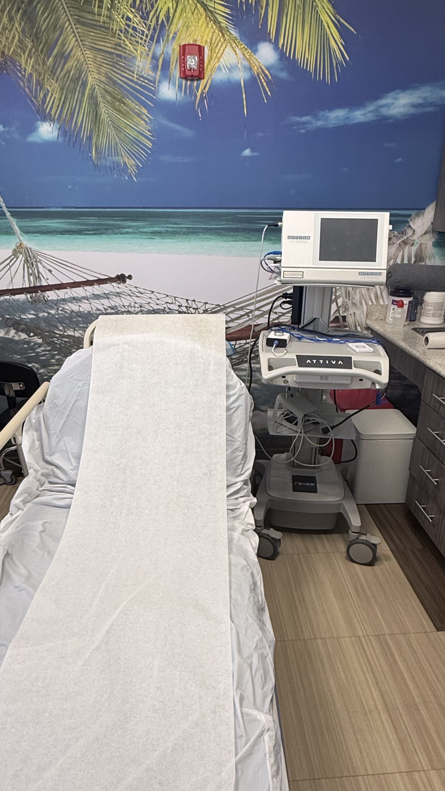
We’ve compiled answers to the most common questions our clients ask to help you get a better understanding of how we can assist you.
Elastic Scattering Spectroscopy (ESS) is a form of sub-diffuse reflectance spectroscopy that detects disease-associated changes at the cellular and subcellular level by analyzing how light interacts with tissue architecture. Unlike taking images of lesions, ESS translates tissue morphology directly into spectral features that correlate with histopathologic characteristics. ESS has been evaluated with 30+ peer-reviewed publications on clinical studies that show its ability to distinguish malignant from benign tissue across different tissue types.
DermaSensor delivers short pulses (~30 microseconds) of broadband white light spanning 300-900 nm wavelengths. When this light penetrates tissue, photons scatter from refractive-index gradients associated with micro- and nano-scale structures, including nuclear size, chromatin condensation, cellular architecture, and collagen organization. An adjacent collection fiber captures backscattered photons and conveys them to a microspectrometer.
The system records a spectral signature of backscattered intensity versus wavelength. Malignant transformation alters cellular and subcellular structures, creating distinct optical signatures in the ESS spectrum. Lesions with different morphology, cellular, and subcellular features have different densities. The recorded spectrum undergoes preprocessing (smoothing and downsampling) before analysis by the proprietary neural network machine learning algorithm, which compares the pattern against training data from over 2,000 histopathologically confirmed lesions.
Time-gated detection enables the device to function in ambient room light without requiring darkening, making it practical for clinical workflows. The entire measurement takes seconds and requires no tissue preparation.
Once captured, the spectral data is analyzed by a trained algorithm. The algorithm has been trained and validated using:
Over 20,000 spectral scans, and more than 4,500 skin lesions
Histologically confirmed melanoma, BCC, and SCC
Benign lesions, including unbiopsied lesions diagnosed by board-certified dermatologists